
Organoids: Modeling Humanity
Written by David Kim-Shoemaker
Edited by Claudia Reines
How does one study the human body? Psychologists, anthropologists, and sociologists can much more easily answer this question than biologists can. Humans are expensive to keep, take up significant portions of space, and the inclusion of ethics only makes research exponentially more difficult. It is here that scientists have turned to animals for the advancement of science. From the Greek anatomists Galen and Erasistratus to the most advanced laboratories of today, laboratory animals have been invaluable.
However, animals are not perfect models of humans. As important as they were for the establishment of many of biology’s founding principles, they often fall short when considering genetics and molecular biology. This, coupled with the common goal of removing as many variables as possible from an experimental system, led to the need for cell culturing: growing cells from organisms outside of their bodies.
Robert Koch, often considered the father of microbiology, took the first step of many in accomplishing such a task. Through placing liquid growths of bacteria in gelatin (later he would use agar) and cooling the entire system, he was able to create a solid, homogenous, and observable culture medium.
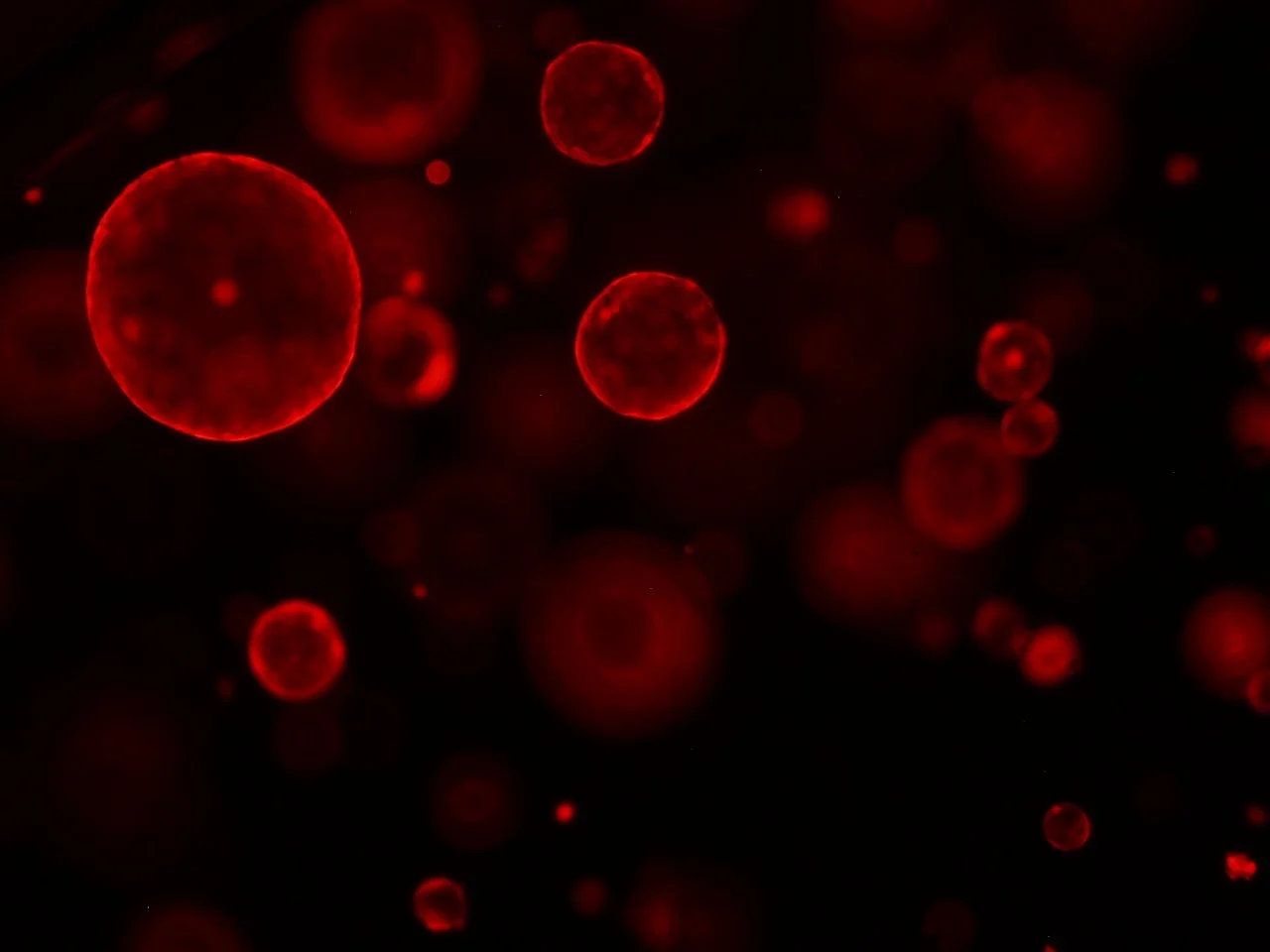
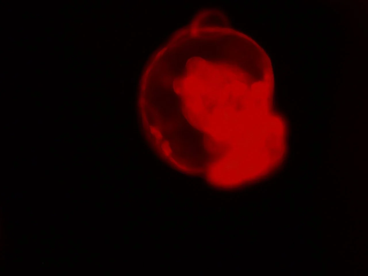

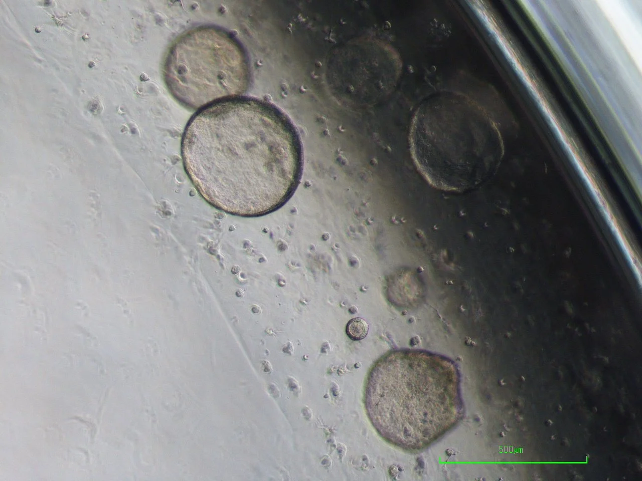
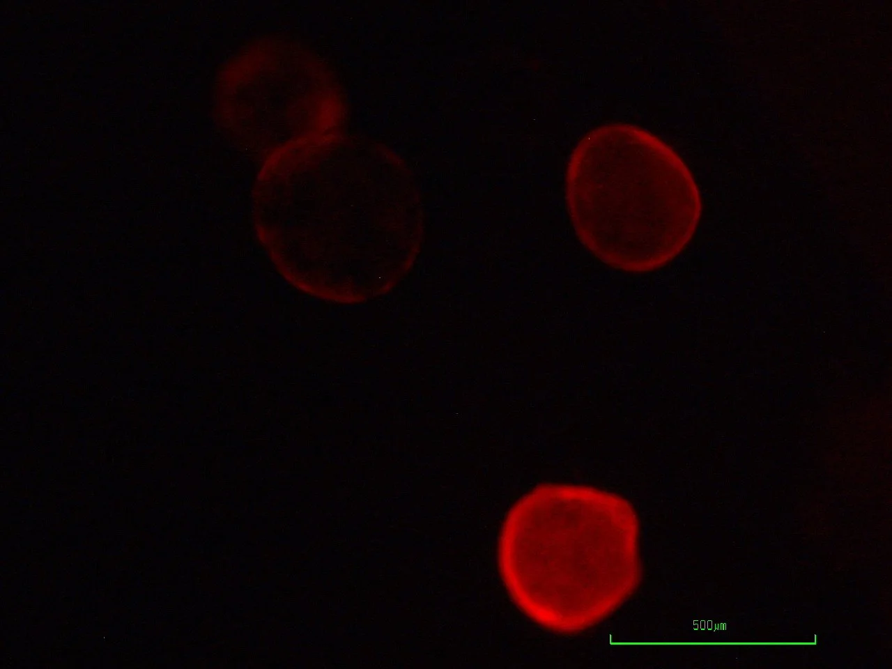
Photos provided by the Nikitin Lab of Cornell University. Curated by EllaRose Sherman (eks92@cornell.edu)
Ross Harrison would later repurpose some of these methods using the hanging-drop technique in 1910. Unlike Koch’s use of bacteria, Harrison grew neurons. Via attachment of a small drop of cells or tissue fragments to an inverted coverslip with a small amount of growth medium, this
created an observable environment where the cells could temporarily grow. Although observable for a short duration, long-term growth was not feasible given the small volume of the drop.
The Carrel flask (from Alexis Carrel) would help resolve this issue by using a flat, round glass vessel with angled necks to help maintain sterile conditions. Given the larger volume, cell cultures could be grown and even transferred over time. More interestingly, Carrel and his successors used this method to maintain a line of chick heart tissue for thirty-four years, which led to the idea that cells could be grown outside of the body for an infinite duration. Today it is speculated that such growth of “normal” cells could not be possible without any experimental issues, as most cells will die after repeated cycles of growth due to the Hayflick limit. But it was nonetheless evidence that cells could be maintained long-term in laboratory conditions. The greatest example of this, HeLa, was found to have truly limitless replication. To this day, it remains used in scientific research.
The next question became the organization of tissues in laboratory conditions. All previously mentioned methods are considered two-dimensional (2D), as cells only expand in longitudinal and lateral directions when placed on a flat surface. After all, the most defining trait of multi cellular organisms is that they are organized and work in concert.

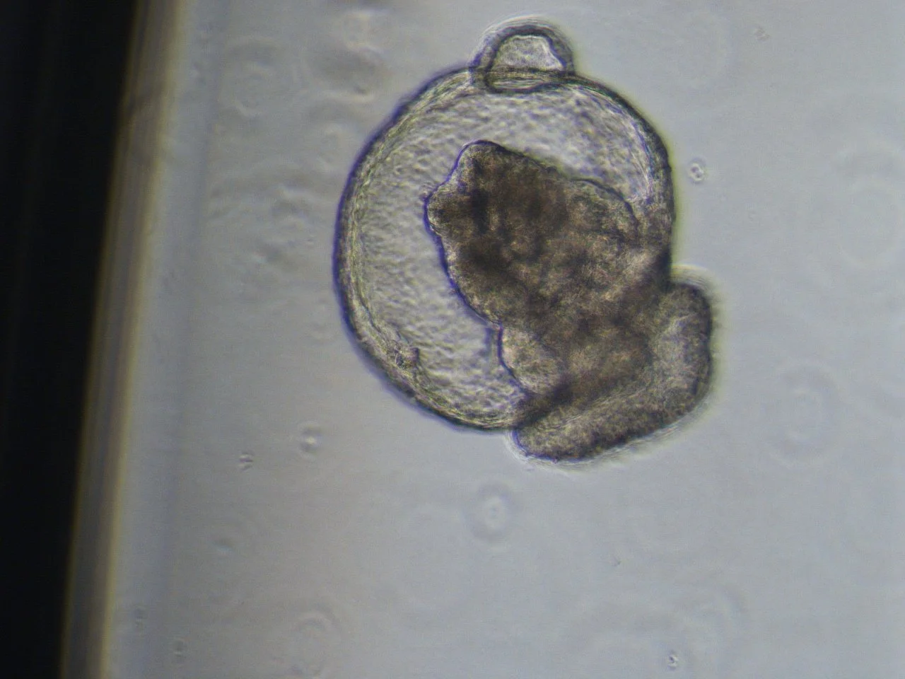
Photos provided by the Nikitin Lab of Cornell University. Curated by EllaRose Sherman (eks92@cornell.edu)
Adopting a scaffold in cultures would allow for a greater extent of three-dimensional (3D) cell growth. Matrigel, a matrix from sarcoma cells, was similar to the extracellular environment where many animal cells proliferate. Spheroid cultures could be used to create cells that clumped together, meaning they themselves acted as a scaffold for other cells. Such methods could be used to produce organoids, models for organs.
Again, these are not exactly accurate representations of entire body systems. Organs are connected to each other, maintained, and developed via signals from the vascular system. This would lead to the organ-on-a-chip system: an extremely advanced composition of microtubes and structures designed to mimic the delivery of nutrients and signals throughout an organ.
It is doubtful that our advancement will end with organ-on-a-chip technologies, the model that comes after it, or even the model after its successor. Over two thousand years after the scientific work of the Greek anatomists, we still struggle to understand ourselves: a testament to our innate complexities and desire for continuous improvement.
David Kim-Shoemaker ‘29 is in the College of Agriculture and Life Sciences. He can be reached at djk323@cornell.edu.